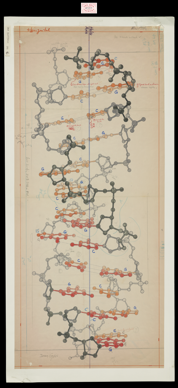
Z-DNA
1983, 34 3/8" x 15 1/8"

In this sketch, Geis illustrates the left handed Z-form of double stranded deoxyribonucleic acid (DNA). Z-DNA is indicated by the zig-zag like pattern of the two strands in relationship to each other. Geis shows a line of symmetry down the middle of the illustration highlighting the helix axis of the molecule.
Used with permission from the Howard Hughes Medical Institute (www.hhmi.org). All rights reserved.

Related PDB Entry: 2DCG
Experimental Structure Citation
Wang, AH, Quigley, GJ, Kolpak, FJ, Crawford, JL, van Boom, JH, van der Marel, G., & Rich, A. (1989). Molecular Structure Of A Left-Handed Double Helical Dna Fragment At Atomic Resolution. Nature, 282, 680-686. doi:10.2210/pdb2dcg/pdb
About Z-DNA
Z-DNA is a duplex structure, with the two strands of the molecule coiling in left-handed helices and a pronounced zig-zag pattern in the phosphorous backbone. Z-DNA can form more easily in an alternating purine-pyrimidine sequence , which is what leads to the zig-zag pattern.


