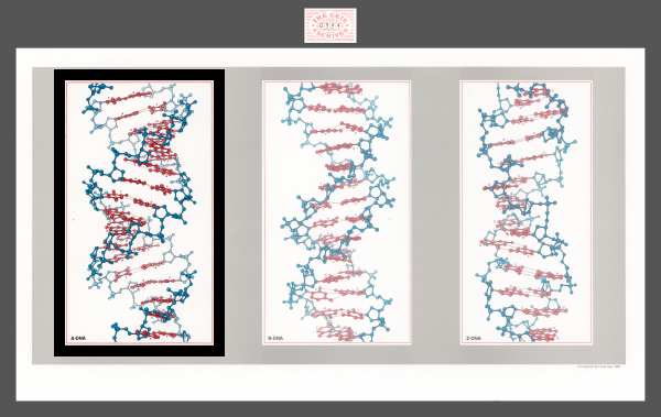
A-DNA
1983
Geis’ depicted version of the A-helix is different from the B-helix. The bases are slightly more offset and the major groove is deeper.


Used with permission from the Howard Hughes Medical Institute (www.hhmi.org). All rights reserved.

Related PDB Entry: 1ANA
Experimental Structure Citation
Conner, B. N., Yoon, C., Dickerson, J. L., & Dickerson, R. E. (1984). Helix geometry and hydration in an A-DNA tetramer: IC-C-G-G. J. Mol. Biol. 174, 663-695.
About A-DNA
Molecule of the Month: A-DNA
The DNA A-helix exists under dehydrated conditions (Goodsell, 2001). It is right-handed and consists of 11 base pairs per turn ("B-Form, A-Form, Z-Form of DNA" website). In comparison to the B-helix (most common form of DNA), it has tipped bases and a deep major groove (Goodsell, 2001).
Note: The depicted Chimera and Jmol structures (consisting of 12 base pairs) are predictions by Dr. Wilma Olsen based on the crystallized fragment (consisting of 4 base pairs) to match Geis’ illustration (Zheng et al., 2009).
Text References
B-Form, A-Form, Z-Form of DNA. University of California, Davis. URL: http://biowiki.ucdavis.edu/Core/Genetics/Unit_I%3A_Genes%2C_Nucleic_Acids%2C_Genomes_and_Chromosomes/2%3A_Structures_of_nucleic_acids/B-Form%2C_A-Form%2C_Z-Form_of_DNA
Goodsell, D. (2001). Molecule of the Month: DNA. DOI: 10.2210/rcsb_pdb/mom_2001_11
Zheng G., Lu X. J., & Olson W. K. (2009). Web 3DNA—a web server for the analysis, reconstruction, and visualization of three-dimensional nucleic-acid structures. Nuc. Acids Res. 37 (Web Server Issue), W240-W246
