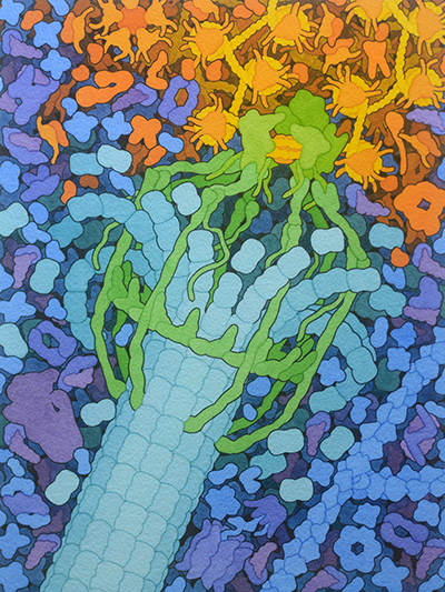
Molecular Landscapes by David S. Goodsell
Kinetochore, 2024
Acknowledgement: Illustration by David S. Goodsell, RCSB Protein Data Bank and the Scripps Research Institute. doi: 10.2210/rcsb_pdb/goodsell-gallery-050
Kinetochores separate chromosomes when eukaryotic cells divide. This illustration shows a yeast kinetochore (green) attached to a disassembling microtubule (blue). Chromatin is shown at upper right with DNA in yellow and histones in orange.


