Molecular Landscapes by David S. Goodsell
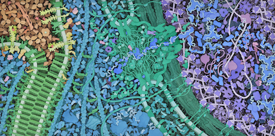
Vascular Endothelial Growth Factor (VegF) Signaling, 2011
Acknowledgement: Illustration by David S. Goodsell, RCSB Protein Data Bank. doi: 10.2210/rcsb_pdb/goodsell-gallery-041
This painting depicts the VegF signaling pathway and some of its consequences. Blood serum is at upper left, with VegF in red. Cell membranes are shown in green at left, with VegF receptor in yellow green near the top and a disassembling adherens junction in darker green at bottom. Multiple kinases (in pink inside the cell) are activated and travel through the nuclear pore (green, at center) to phosphorylate transcription factors in the nucleus (at right).
The painting was created as part of the “Connecting Researchers, Educators and Students” project at the Center for BioMolecular Modeling at MSOE. More information on the project and keys for the painting are available at the CREST site and in the BAMBED publication describing the project:
Span, Elise A., Goodsell, David S., Ramchandran, Ramani, Franzen, Margaret A., Herman, Tim and Sem, Daniel S. 2013. Protein Structure in Context: The Molecular Landscape of Angiogenesis. Biochemisty and Molecular Biology Education 41(4):213-223.


