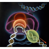
Protein Synthesis
building the molecules of life
Each cell includes a DNA genome that encodes the instructions for building all of the cell's proteins. Atomic structures have revealed many of the molecular machines that protect and replicate this DNA, transcribe it into RNA, and translate it into proteins.
Molecule of the Month Articles (55)
 |
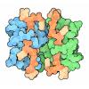
AAA+ Proteases
AAA+ proteases are ATP-powered molecular motors that thread protein chains through a hole |
 |
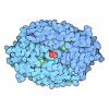
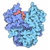
Aconitase and Iron Regulatory Protein 1
Aconitase performs a reaction in the citric acid cycle, and moonlights as a regulatory protein |
 |
Adenine Riboswitch in Action
XFEL serial crystallography reveals what happens when adenine binds to a riboswitch |
 |
Aminoacyl-tRNA Synthetases
Aminoacyl-tRNA synthetases ensure that the proper amino acids are used to build proteins |
 |
Cascade and CRISPR
Cascade and CRISPR help bacteria remember how to fight viral infection |
 |
Catabolite Activator Protein
CAP senses the level of sugar and mobilizes the proteins needed to utilize it |
 |
Chaperones
Chaperones help new proteins fold into their proper shape |
 |
DNA
Atomic structures reveal how the iconic double helix encodes genomic information |
 |
DNA Helicase
DNA helicase pries apart the two strands in a DNA double helix, powered by ATP |
 |
DNA Ligase
DNA ligase reconnects broken DNA strands, and is used to engineer recombinant DNA |
 |
DNA Methyltransferases
Cells add methyl groups to their DNA to encode additional epigenetic information |
 |
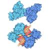
DNA Polymerase
DNA polymerase makes an accurate copy of the cell's genome |
 |
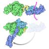
Elongation Factors
Protein synthesis requires the assistance of several elongation factors that guide each step |
 |
Enhanceosome
Enhanceosomes help decide the appropriate time to transcribe a gene |
 |
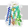
Estrogen Receptor
Estrogen binds to receptors in the nucleus and affects key genes in development |
 |
Exosomes
Exosomes destroy messenger RNA molecules after they have finished their jobs |
 |
Expressome
In bacteria, ribosomes start building proteins as messenger RNA is being transcribed |
 |
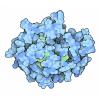
Hemoglobin
Hemoglobin uses a change in shape to increase the efficiency of oxygen transport |
 |
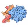
HIV Reverse Transcriptase
HIV builds a DNA copy of its RNA genome, providing a unique target for drug therapy |
 |
Hsp90
Heat shock proteins ensure that proteins remain folded and active under harsh conditions |
 |
Initiation Factor eIF4E
Initiation factors for protein synthesis interact through disordered chains. |
 |
Inteins
Inteins splice themselves out of larger protein chains |
 |
lac Repressor
A genetic circuit controls the production of lactose-utilizing enzymes in bacteria |
 |
Lysozyme
Lysozyme attacks the cell walls of bacteria |
 |
Mediator
Mediator integrates regulatory information to decide when genes need to be transcribed. |
 |
Messenger RNA Capping
Messenger RNA molecules are capped with an inverted nucleotide |
 |
Nucleosome
The cell's genome is stored and protected by nucleosomes |
| O-GlcNAc Transferase
Some protein functions are regulated when sugars are attached |
|
 |
Oct and Sox Transcription Factors
Transcription factors decide when particular genes will be transcribed |
 |
Oligosaccharyltransferase
Oligosaccharyltransferase adds a protective coat of carbohydrates to proteins. |
 |
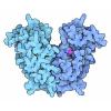
p53 Tumor Suppressor
p53 tumor suppressor protects the body from DNA damage and cancer |
 |
Poly(A) Polymerase
Poly(A) polymerase adds a long tail of adenine nucleotides at the end of messenger RNA |
 |
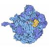
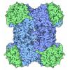
Proteasome
Proteasomes destroy damaged or obsolete proteins inside cells |
 |
RecA and Rad51
Broken DNA strands may be repaired by matching sequences in a duplicate copy of the DNA |
 |
Restriction Enzymes
Bacterial enzymes that cut DNA are useful tools for genetic engineering |
 |
Rhomboid Protease GlpG
Some proteases cut proteins embedded in cell membranes |
 |
Ribonuclease P
The ribozyme ribonuclease P cleaves pre-tRNA to form functional tRNA. |
 |
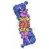
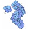
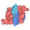
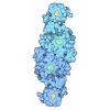
Ribosomal Subunits
Atomic structures of the ribosomal subunits reveal a central role for RNA in protein synthesis |
 |
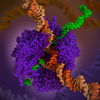
Ribosome
Ribosomes are complex molecular machines that build proteins |
 |
Ribosome Diversity
By comparing the structures of ribosomes from different organisms, we can explore the evolution of life. |
 |
Riboswitches
Special sequences of messenger RNA can bind to regulatory molecules and affect synthesis of proteins |
 |
RNA Polymerase
RNA polymerase transcribes genetic information from DNA into RNA |
 |
Selenocysteine Synthase
Selenium is used in place of sulfur to build proteins for special tasks |
 |
Self-splicing RNA
Special sequences of RNA are able to splice themselves |
 |
Sirtuins
Sirtuin activation is being explored as a way to slow aging. |
 |
Sliding Clamps
Sliding clamps slide along DNA strands and keep DNA polymerase on track during replication |
 |
Small Interfering RNA (siRNA)
Our cells continually look for pieces of double-stranded RNA, a possible sign of viral infection |
 |
TATA-Binding Protein
TATA protein tells RNA polymerase where to get started on a gene |
 |
Thymine Dimers
Ultraviolet light damages our DNA, but our cells have ways to correct the damage |
 |
Topoisomerases
Topoisomerases untangle and reduce the tension of DNA strands in the cell |
 |
Transfer RNA
Transfer RNA translates the language of the genome into the language of proteins |
| Transfer-Messenger RNA
tmRNA rescues ribosomes that are stalled by damaged messenger RNA |
|
 |
Transposase
Transposases shift genes around in the genome |
 |
Ubiquitin
Ubiquitin is used to tag obsolete proteins for destruction |
 |
Zinc Fingers
Zinc ions are used to strengthen small protein modules that recognize DNA |
Learning Resources (12)
 |
DNA
Paper Model
Atomic structures reveal how the iconic double helix encodes genomic information
|
 |
Paper Nucleic Acid Models for Hands-on Education
Paper Model
Download templates for a set of paper models that can be cut from paper or card stock and used for demonstrations of the fundamentals of DNA structure, DNA replication, and RNA translation.
|
| tRNA
Paper Model
Transfer RNA translates the language of the genome into the language of proteins
|
|
 |
ADN: El Acido Desoxirribonucleico (Spanish)
Flyer
Atomic structures reveal how the iconic double helix encodes genomic information
|
 |
Award-winning RNA Polymerase Illustration
Poster
|
 |
The Ribosome
Flyer
This flyer commemorates the 2009 Nobel Prize in Chemistry for studies of the structure and function of the ribosome.
|
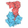
| Shiga Toxin2 in Complex with Ribosomal P-stalk
Poster
Cryo-EM structure of Shiga toxin 2 in complex with the native ribosomal P-stalk reveals residues involved in the binding interaction.
|
|
 |
Ribosomal Subunits
GIF
Atomic structures of the ribosomal subunits reveal a central role for RNA in protein synthesis. Ribosomes are complex molecular machines that build proteins.
|
 |
The Biologist Magazine Big Biochemical Colouring-in Series
Coloring Book
This series was created for The Biologist in 2022 to invite readers to get artistic while learning about some of the life sciences’ most important and interesting macromolecules.
|
| Commemorating 75 Years of Discovery and Innovation at the NSF
Other Resource
Download images celebrating NSF and PDB milestones
|
|
| Molecular Backgrounds For Virtual Meetings
Other Resources
Download images created by David Goodsell to add a molecular backdrop to your next virtual meeting. Click on the image to expand.
|
|
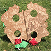 |
Hemoglobin Bean Bag Toss
Other Resource
|
Curriculum Resources (3)


Structural Biology Highlights (20)


Geis Digital Archive (7)
| Transfer Ribonucleic Acid (tRNA)
Geis illustrates the structure of transfer ribonucleic acid (tRNA), the nucleic acid that translates the language of the genome into the language of proteins. |
|
 |
Deoxyribonucleic Acid (DNA)
Geis illustrates three possible forms of deoxyribonucleic acid (DNA). He highlights the differences between each structure by displaying them in a side-by-side manner. |
 |
B-DNA
Geis illustrates B-DNA in blue looking from above, through the double helix. The two bases on top are highlighted in white to distinguish one individual section of the layered scene. |
 |
DNA
Geis illustrates a double helix in his depiction of DNA. He portrays the helices with a soft ribbon structure. The white "box-like" structures represent a base pair in the DNA strand.
|
 |
A-DNA
Geis uses a thin ball and stick representation of a section of A-DNA, the more compact conformation of DNA less often seen in biological systems. He draws it from a perspective looking down into the double helix, showing the increase in diameter of the middle of the helix from the B form DNA.
|
 |
Z-DNA
In this sketch, Geis illustrates the left handed Z-form of double stranded deoxyribonucleic acid (DNA). Z-DNA is indicated by the zig-zag like pattern of the two strands in relationship to each other. Geis shows a line of symmetry down the middle of the illustration highlighting the helix axis of the molecule.
|
 |
TATA-Binding Protein (TBP)
Geis visualizes the TATA-box binding protein (TBP) and its associated proteins that form a preinitiation complex in all eukaryotes for transcribing DNA to messenger RNA. Acrylic painting in collaboration with Dr. Stephen K. Burley (Howard Hughes Medical Institute and Rockefeller University). Illustration copyright by Irving Geis. |
Goodsell Molecular Landscapes (8)
 |
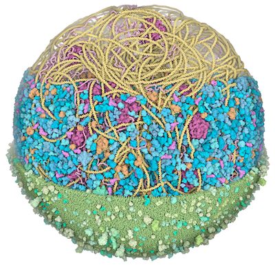
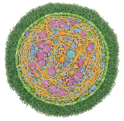
JCVI-syn3A Minimal Cell
Cross-section through an entire JCVI-syn3A minimal cell showing an entire cellular proteome.
|
 |
RecA and DNA
RecA assists with the pairing of DNA strands during DNA repair.
|
 |
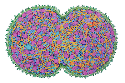
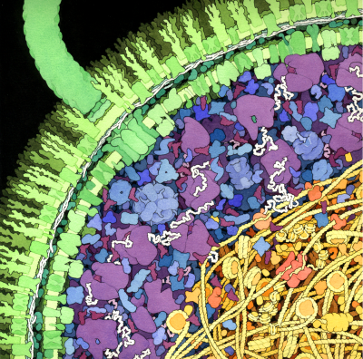
Model of a Mycoplasma Cell
Image of a 3D model of an entire mycoplasma cell.
|
 |
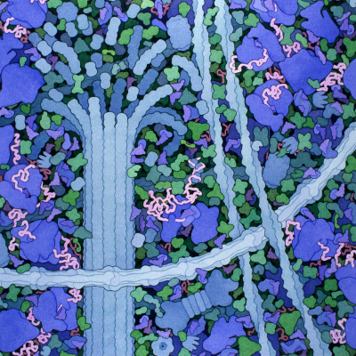
Escherichia coli Bacterium
A cross-section through an Escherichia coli cell reveals the crowded nature of the cell and diverse molecular processes.
|
 |
Mycoplasma mycoides
Mycoplasma mycoides (2011) by David S. Goodsell. doi: 10.2210/rcsb_pdb/goodsell-gallery-011
|
 |
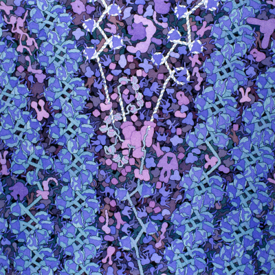
Biosites: Cytoplasm
Biosites: Cytoplasm (2005) by David S. Goodsell
|
 |
Biosites: Nucleus
Biosites: Nucleus (2005) by David S. Goodsell
|
 |
Escherichia coli
Escherichia coli (1999) by David S. Goodsell
|
